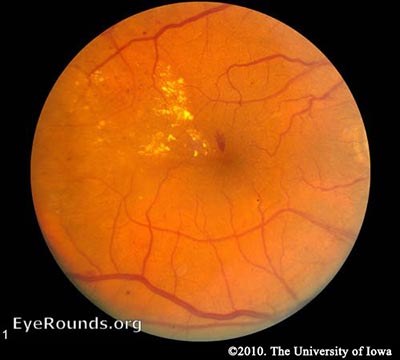
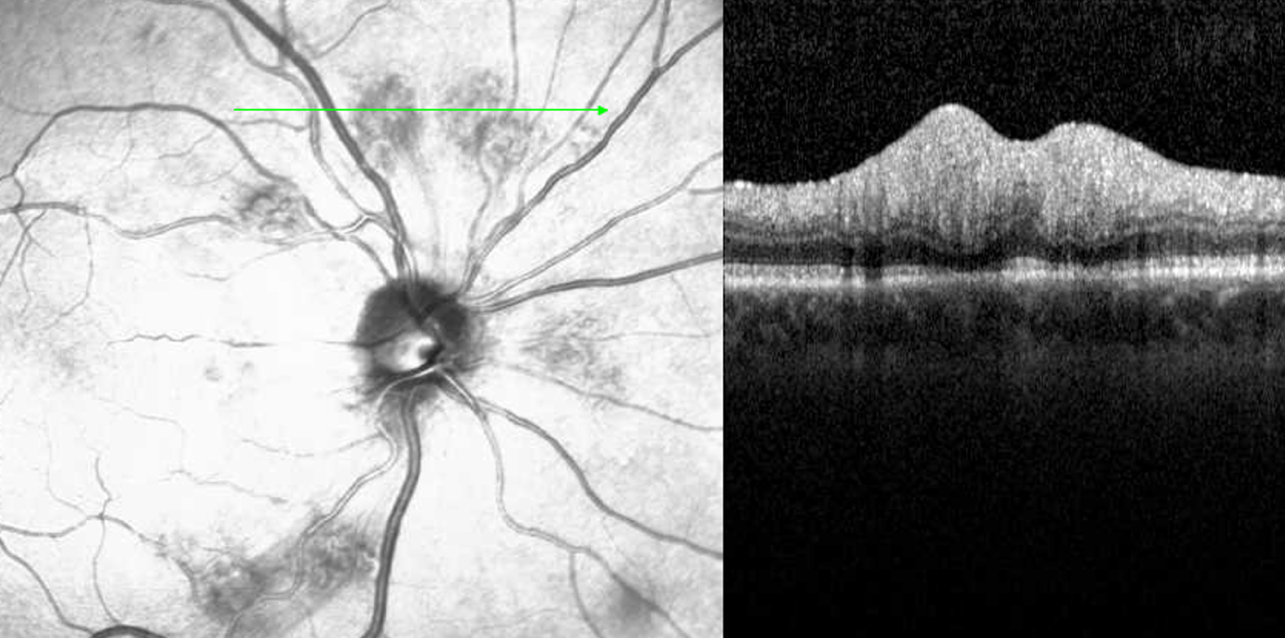
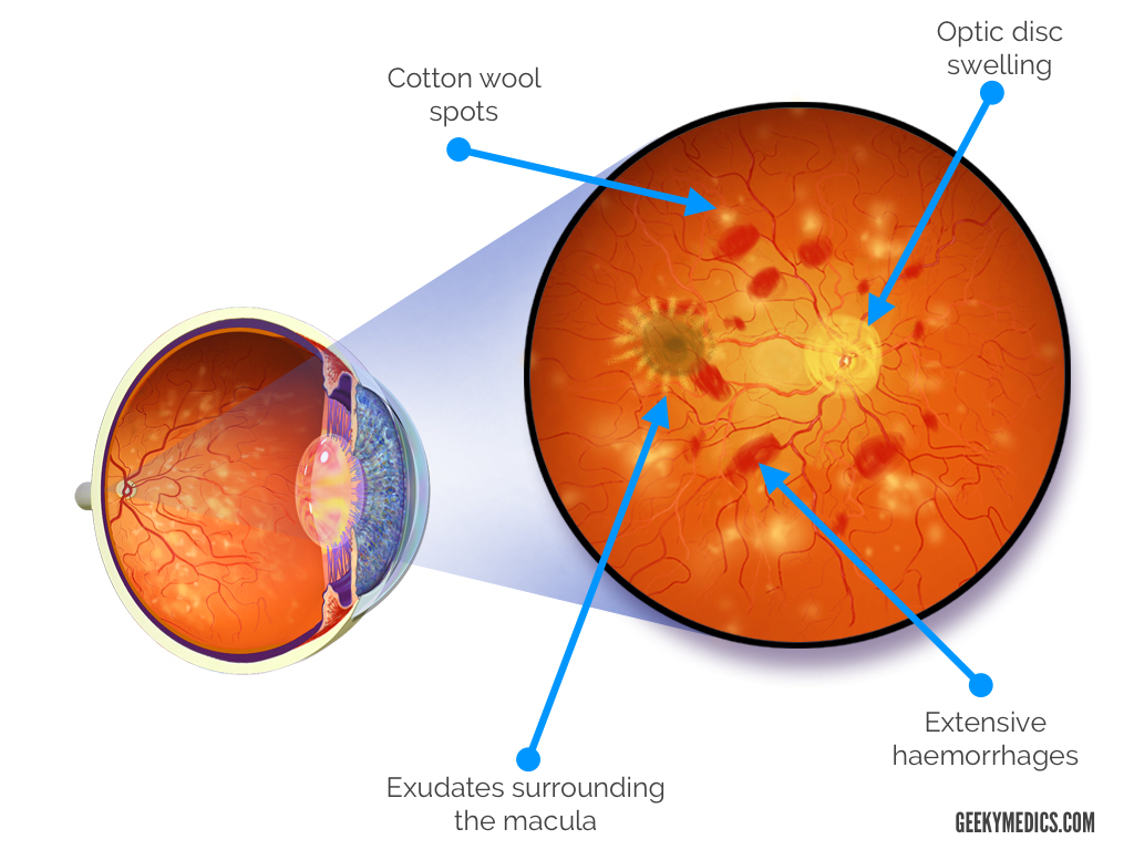
Cotton Wool Spots : Ophthalmoscopic Abnormalities : The Eyes Have It
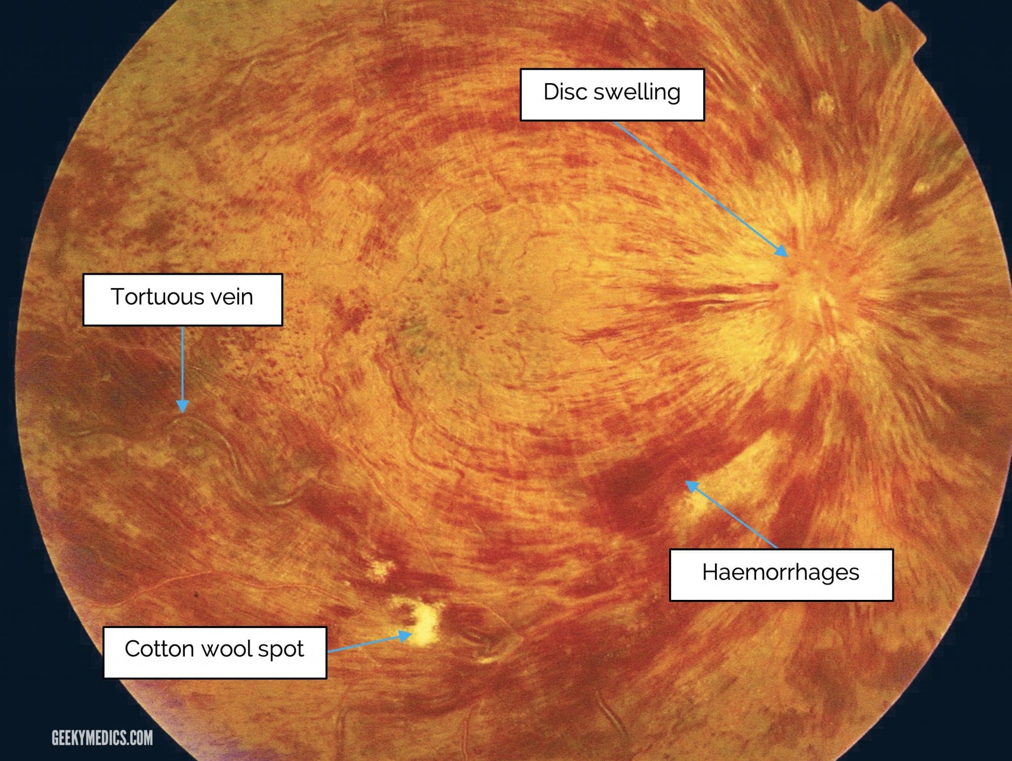
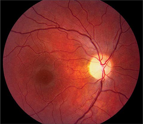
Fundoscopic Appearances of Retinal Pathologies
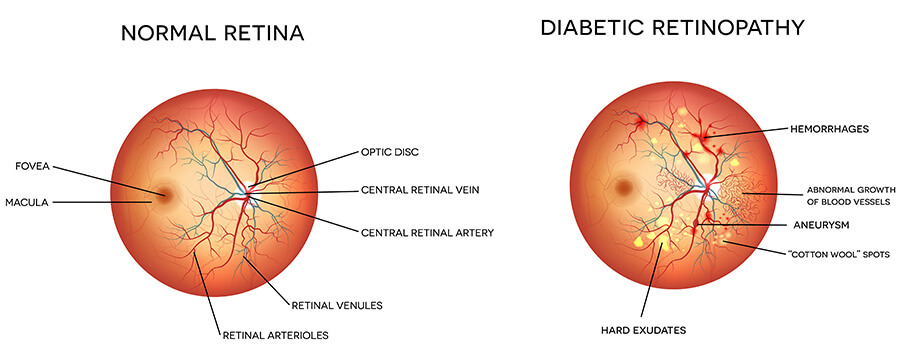
Diabetic Retinopathy for Medical Students. Classification

Retinal Vasculopathy With Cerebral Leukoencephalopathy and Systemic Manifestations: Critical Role of Retina Specialists - Odette M. Houghton, Jonathan Carter, Radhika Dhamija, 2023
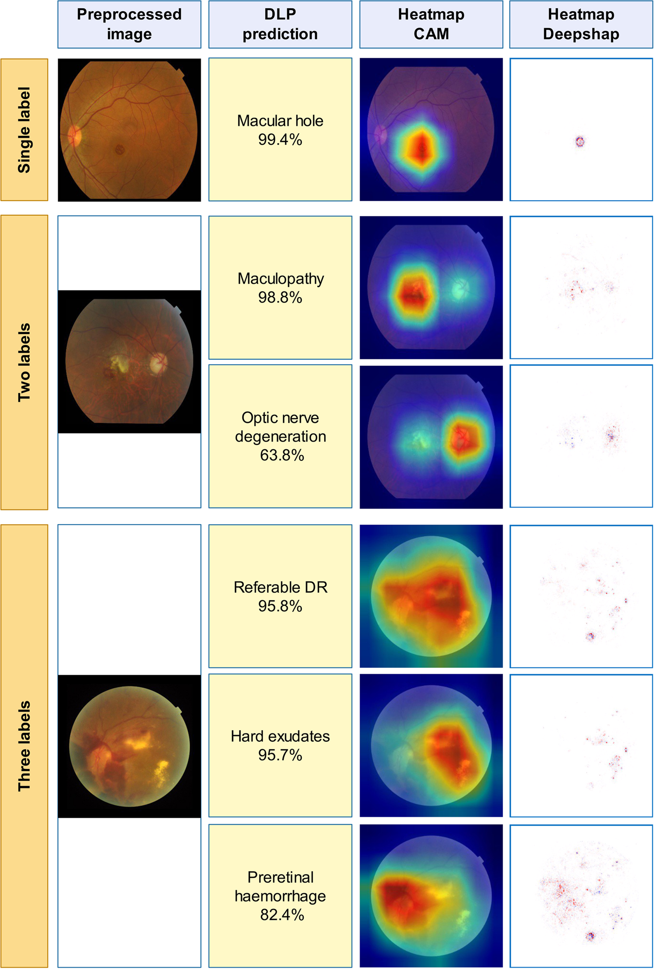
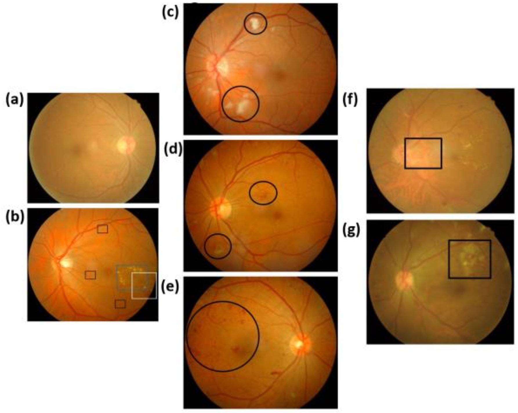
Automatic detection of 39 fundus diseases and conditions in retinal photographs using deep neural networks
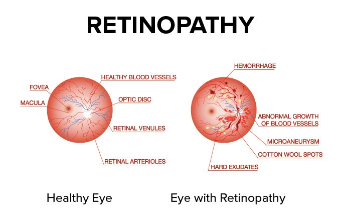
Retinopathy - All About Vision
Cotton Wool Spots in a Patient with COVID-19 Published in CRO (Clinical & Refractive Optometry) Journal

Cotton Wool Spots - an overview
JCM, Free Full-Text
Lesson: Can You Spot These Retinal Vascular Abnormalities?
Fundoscopic Appearances of Retinal Pathologies
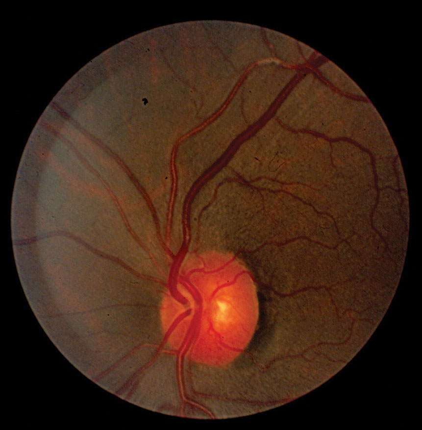
Use of the Hand-Held Ophthalmoscope
Branch Retinal Artery Occlusion : Ophthalmoscopic Abnormalities : The Eyes Have It

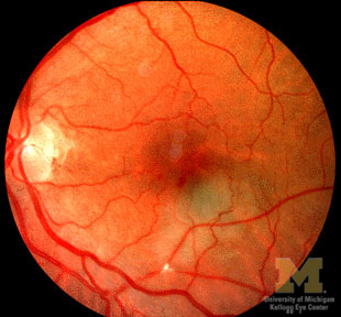
a Right eye fundoscopy showing cotton wool spots following the path of

8 Arterial Occlusive Disease
Diabetic Eye Care - Richard Manganiello, MD